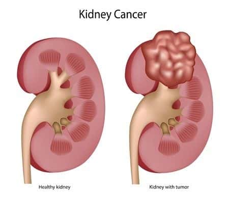
A family medicine expert witness opines on a case involving a fifty-five-year-old female with a past medical history of migraines and IBD. She presented to her primary care physician complaining of non-specific pain in the abdomen and back. On examination of the patient, the treating physician did not find anything significant. The physician ordered a CT scan of the abdomen and pelvis. The scan revealed a sub-centimeter hyper-dense lesion involving the left mid kidney that may have represented a hyper-dense cyst, or other etiology. The radiologist who reported these findings suggested to the treating physician that follow up with a repeat sonogram or CT scan in three months was warranted to monitor the mass. The treating physician did not order any further investigation of the mass. The physician did not order follow up of the patient. Sometime later, the patient returned with severe lower back pain, abdominal pain, and lower right-sided pain. Physical examination of the patient’s abdomen revealed a palpable mass in the right flank. Upon repeat CT scan of the abdomen and pelvis, it was the revealed that the mass, which was less than a centimeter on initial medical imaging, had grown to four centimeters.
Question(s) For Expert Witness
1. What is the protocol for following up with renal masses seen on CT?
2. What is the prognosis of renal cell carcinoma diagnosed at an early stage?
Expert Witness Response
Most instances of renal cell carcinoma are found incidentally while imaging the abdomen for unrelated problems. Because most solid masses of the kidney are ultimately found to be cancerous, further work-up of these masses is necessary. Since lesions that are less than a centimeter are difficult to sample and have a low risk of metastasizing, it is best to follow these lesions with serial imaging, such as with repeat CT scans. Typically this would be done in 3 to 6 month intervals for a year, and then annually. If the mass were to grow large enough, biopsy or surgical removal would be ideal. Early detection of renal cell carcinoma has an excellent prognosis, with an approximate 5-year survival rate of 90% when detected at Stage I. However, survival decreases as the tumor size increases or begins invading the surrounding tissue. By the time Stage IV involvement is reached, 5-year survival plunges to less than 10%. Clearly, failure to follow the progression of a small, solid renal mass can have a significant impact on the likelihood of survival from renal cell carcinoma, and failure to follow the radiologist's recommendations on serial imaging in this particular case likely allowed the renal mass to progress before a diagnosis could be made and appropriate treatment could be instituted.
About the author
Dr. Faiza Jibril
Dr. Faiza Jibril has extensive clinical experience ranging from primary care in the United Kingdom, to pediatrics and child abuse prevention at Mount Sinai Hospital, to obstetrics in Cape Town, South Africa. Her post-graduate education centered on clinical research and medical ethics. Dr. Jibril is currently Head of Sales in the US and Canada for Chambers and Partners - a world leading legal ranking and insights intelligence company.


